

-

Telephone
Hot line
-

Phone Call
+86-15221725700
-

Go To Top
Contact us
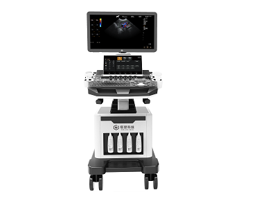
Animal Color Doppler Ultrasound
Product model:
DU T5VDetailed introduction:
Product Name: Animal Color Doppler Ultrasound
Features
1. Color Doppler echocardiography
2. Contains micro- convex probe , linear array probe , phased array probe , 2 1.8 -inch widescreen , 3.3-inch touch screen , channel optimization and improvement , with 4 probe interfaces and PC platform
3. Meet the inspection and diagnosis of pet hospitals and scientific research institutions in digestive system, reproductive system, urinary system, physical examination and other aspects.
Parameter
1. Product Name: Color Doppler Animal Ultrasound Diagnostic Instrument |
1.1 Structure type: double-screen cart type |
2. Instructions for use and cargo requirements 2.1 Meet the inspection and diagnosis of pet hospitals and scientific research institutions in digestive system, reproductive system, urinary system, physical examination and other aspects. |
3. System technical specifications and overview 3.1. Mainframe of full digital color Doppler ultrasound diagnostic system |
3.2. Digital beam booster |
3.3. Multiple beamforming |
3.4. Two-dimensional grayscale mode |
3.5. Harmonic imaging technology |
3.6. B+C dual real-time mode |
3.7. M-mode |
3.8. Anatomical M mode, sampling lines ≥ 3 |
3.9. Doppler imaging (including color, energy, and directional energy Doppler modes) |
3.10. Spectral Doppler imaging (including pulsed Doppler, high pulse repetition frequency, continuous wave Doppler) |
3.11. Tissue Doppler imaging (including tissue velocity map, M-mode, spectral imaging and other modes) |
3.12. Four-dimensional imaging |
3.13. ★Contrast imaging technology (optional) |
3.14. ★With PView wide-view imaging technology (optional) |
3.15. Spatial composite imaging technology (can be used in abdomen, obstetrics, blood vessels, small superficial organs, and can be displayed on the same screen for double contrast) |
3.16. Frequency Composite Imaging |
3.17. Extended imaging |
3.18. Real-time double-contrast imaging |
3.19. Real-time three simultaneous imaging (two-dimensional, color, spectrum real-time simultaneous imaging) |
3.20. Speckle noise suppression technology |
3.21. Elastography technology (optional) |
4. ★ Operating interface: The host includes 10 operating language interfaces . |
5. System technical parameters and requirements 5.1 Standard ≥ 21.5 -inch high-resolution color LCD display (optional 23.8-inch high- resolution color LCD display ) 13.3-inch color LCD touch screen, support multi-touch |
5.2 ★The built-in probe interface of the host is more than 4, fully activated, the size is the same, interoperable |
5.3. 2D Grayscale Mode 1) Digital beamformer 2) Digital full dynamic focusing, digital variable aperture and dynamic apodization, A/D≥1 5 bit 3) Receiving mode: transmit and receive channels ≥ 1024, multiple signals are processed in parallel 4) Scanning line: line density per frame ≥ 512 ultrasonic lines 5) Focusing of emission sound beam: emission ≥ 10 segments, the focus position has a special menu adjustment 6) TGC≥8 segments 7) Gain adjustment: B/M/D are independently adjustable, ≥100dB 8) ★ Dynamic range adjustment: ≥180dB 9) ★Maximum display depth ≥ 360 mm 10) Grayscale: ≥ 6 to 7 levels, visually adjustable 11) Sound power: 1%-100% 12) Two-dimensional independent deflection of linear array probe 13) Partial magnification ( 1.5/2.0/2.5/3.0/3.5, 4.0/4.5/5.0/10 times ) |
5.4. Color Doppler Imaging 1) Imaging method: including speed, speed variance, energy, direction energy display, etc. 2) Display mode: B/C, B/C/M, B/POWER, B/C/PW 3) Linear density ≥ grade 3 4) Color hiding technology: You can hide the color without returning to the 2D mode, and only display the color speed scale 5) Blood flow distribution map function, color blood flow profile to measure intravascular flow velocity |
5.5. Spectral Doppler Mode 1) Display format: full screen, duplex/triple (PW only) 2) Gain: ≥100dB 3) Multi-spectral speed: ≥4 levels adjustable 4) Maximum measurement speed: PWD: positive or reverse blood flow velocity ≥ 7.6m/s; CWD: blood flow velocity ≥ 20.0m/s, minimum velocity: ≤ 5 mm/s (non-noise signal); 5) Zero movement: ≥8 grades 6) Display mode: B, PW, B/PW, B/C/PW, B/CW, B/C/CW, etc. 7) Spectrum automatic measurement, manual measurement 8 ) Display control: inversion, zero shift, B refresh, D extension, B/D extension, etc. 9) Intelligent Doppler technology, which can be freely switched between real-time B+CFM mode and real-time PW mode. |
5.6. ★Standard wide-view imaging (optional) 1) Display length up to 50 cm in high resolution 2) With two-dimensional wide-view and color wide-view imaging modes |
5.7. Probe interface ≥ 4 |
5.8. Probe: broadband frequency conversion probe, two-dimensional and color independent frequency conversion, 1) Probe fundamental frequency conversion number ≥ 9 segments; 2) 1. Convex array probe : fundamental frequency: 2.0MHz/2.3MHz/2.5MHz/3.0MHz/3.5MHz/4.0MHz/4.5MHz/5.0MHz/5.5MHz, nine-segment frequency conversion (depth 50-360mm) 2. Linear array probe : Fundamental frequency: 4.0MHz/4.5MHz/5.0MHz/6.0MHz/7.5MHz/8.0MHz/9.2MHz/10.0MHz/12.0MHz/14.0MHz, nine-segment frequency conversion (depth 20-120mm) 3. Phased array probe : Fundamental frequency: 1.7MHz/1.9MHz/2.0MHz/2.5MHz/3.0MHz/3.4MHz/3.8MHz/4.2MHz/5.5MHz, nine-segment frequency conversion (50-360mm) Harmonic frequency: 3.0MHz/4.0MHz/5.0MHz, three-stage frequency conversion 4. Micro-convex probe : Fundamental frequency: 3.0MHz/3.5MHz/4.0MHz/5.0MHz/5.4MHz/6.0MHz/7.0MHz/8.0MHz, eight-segment frequency conversion (depth 30-160mm) Harmonic frequency: 6.0MHz/7.0MHz/8.0MHz, three-stage frequency conversion 5. High frequency phased array probe: Fundamental frequency : 3.0MHz / 3.5MHz/4MHz/5MHz/5.4MHz/6MHz/7MHz/8MHz, eight-segment frequency conversion (depth 50-360mm) Harmonic frequency: 6MHz/7MHz/8MHz, three-stage frequency conversion |
6. Measurement/Analysis and Reporting 6.1 Routine measurements: distance, area, ellipse, reticle, angle, distance ratio, volume, volume (ellipse), area ratio, diameter, joint angle. |
6.2 Specialty Measurements 1)Cardiac measurements: LV, MPAD, RVEDd, RVEDs, LV myocardium, LAVol, RV/LV, LVSimps, LA/AO, MV, LVMassA/L, QP/QS. 2) ★Obstetric measurement: Canine: head-rump diameter, fetal sac, transverse cranial diameter, body cavity transverse diameter; Feline: body cavity transverse diameter, cranial transverse diameter; Porcine: pig heart, pig stomach; Bovine: head-rump diameter , trunk transverse diameter, cranial transverse diameter; sheep family: head-rump diameter, biparietal diameter; equine family: fetal sac (H), fetal sac (V) |
7. Peripheral part 7.1. Configure a set of ultrasound graphics workstation, the workstation software needs to have a registration certificate, supports digital black and white, analog black and white, digital color, analog color, text and video printers, and supports foot switches. 7.2 Support network connection 7.3 Support DICOM 3.0 DICOM Obstetrics and Gynecology, Cardiac, Vascular Report 7.4 Video/audio input and output 7.5 Host comes with USB interface 7.6 Support the ultrasound system to send clinical pictures and reports directly to the computer through the network 7.7 Optional couplant heater |
8 Movie playback and raw data processing 8.1 Movie playback ≥3061 frames, support manual and automatic playback 8.2 The capacity of the digital hard disk is ≥120G+1T, which can permanently store dynamic and static images, and can read, transfer and delete images at any time 8.3 Multiple export image formats: dynamic images and static images are directly exported in PC format, and images can be viewed directly on ordinary PCs without special software. While exporting and backing up image data, real-time inspection can be performed without affecting inspection operations 8.4 Support DVD R/W burner 8.5 With professional probe placement racks ≥ 5 (excluding couplant placement racks), each probe placement rack can be replaced left and right |
9 After sales 10.1 Whole machine warranty ≥ 2 years 10.2 Lifetime maintenance after the expiration of the warranty period, and free upgrade and maintenance of the workstation software involved for life 10.3 Regularly conduct free tour maintenance for users 10.4 Repair the fault within 24 hours, if it cannot be solved within 24 hours, provide a backup machine |

 2022-06-16
 hits
hits


