

-

Telephone
Hot line
-

Phone Call
+86-15221725700
-

Go To Top
Contact us
Animal Color Doppler Ultrasound
Product model:
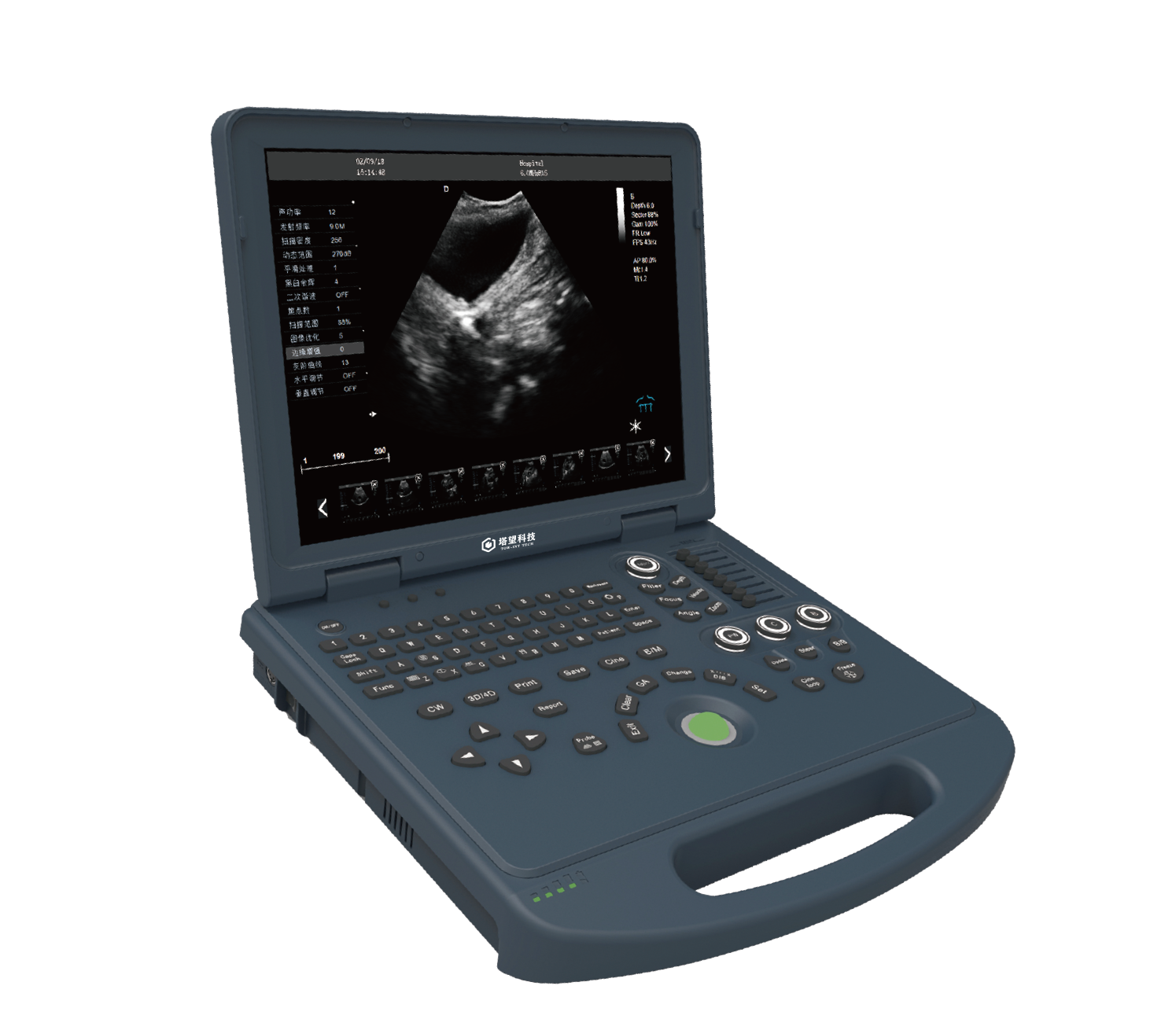
SONA L3VDetailed introduction:
Product Name: animal mobile color ultrasound
Features
1. Adopt intelligent image processing technology , tissue harmonic imaging technology , noise suppression speckle technology , multi-beam parallel processing technology, etc.
2. High-efficiency all-digital image management system, more convenient and flexible configuration for image storage and reading, easy to carry, ergonomic design, greatly improving the scope of use 15-inch high-definition LED display, 180-degree full viewing angle display LED-backlit silicone keyboard, easy to use Darkroom operation Built-in rechargeable lithium battery to cope with various environments
3. Examination and pathological diagnosis of liver, gallbladder, spleen, kidney, bladder, uterus, pregnancy and other tissues and organs of small and medium animals.Pregnancy diagnosis in reptiles, diagnosis in fish sexing
4. Pregnancy examination and backfat thickness measurement, hip fat and muscle measurement of large animals such as cattle, sheep, horses, pigs, camels, etc.
Technical Parameter
1. Product Name: Animal Color Doppler Ultrasound Diagnostic Instrument |
1.1 Structure type: notebook type |
2. Instructions for use and cargo requirements 2.1 It is suitable for ultrasound examination of various needs such as pet hospitals, clinics, zoos, breeding/breeding bases and various scientific research units. |
3. Main specifications and system overview |
3.1 Ultrasound host operating system: Windows 10 operating system 3.2 Spectral Pulse Doppler 3.3 Directional Energy Doppler 3.4 Real-time triple synchronization 3.5 With spatial composite imaging 3.6 Possess tissue harmonic imaging technology 3.7 2B/4B imaging mode 3.8 System language: Chinese, English, French, Russian and Spanish five languages 3.9 Monitor: ≥ 15 inches 3.10 Integrated clipboard: display the saved images at the bottom of the screen, which can be directly transferred or deleted 3.11 The system has the function of field upgrade 3.12 Preset conditions: For different inspections, preset the inspection conditions to optimize the image, reduce the adjustment during operation, and commonly required external adjustment and combined adjustment 3.1 3 probe interface ≥ 1 3.1 4 With trapezoidal imaging function 3.1 5 with one-click intelligent optimization |
4. Probe: |
Linear array probe: Frequency: 6.0MHz/7.5MHz/8.5MHz/10.0MHz/12.0MHz/ H10.0MHz, six-segment frequency conversion (depth 20-128 MM ) convex probe ( R1 1 ): frequency 4.5MHz/ 5.0MHz/ 6.0MHz/ 6.5MHz/ 7.0MHz/ 9.0MHz /H8.0Mhz , seven-segment frequency conversion (depth 30-111MM) Convex probe: frequency 2.0MHz/ 2.5MHz/3.0MHz/3.5MHz/4.0MHz/ 5.5MHz/ H4.0MHz/H5.0MHz, eight-segment frequency conversion (depth 30 - 255 MM) Rectal probe: frequency 4.0MHz/6.5MHz/ 9.0MHz /H8.0Mhz , four-segment frequency conversion (depth 20-110MM ) |
5 2D imaging modes: |
5.1 Gain: 0-100, step 1 can be adjusted visually 5.2 TGC: 8-segment adjustable 5.3 Dynamic range: 20-280dB 20-level visual adjustable 5.4 Pseudo-color: 0-11 grades, visually adjustable 5.5 sound power: 5%-100%, step by 5%, visually adjustable 5.6 Body position markers ≥ 1 8 kinds 5.7 Maximum number of focal points: 6 focal points, which can be moved all the way 5.8 Grayscale: 0-7 levels are visually adjustable 5.9 Filter: 0-4 5.10 Scanning range: 50%-100% 5.11 Frame correlation: 0-4 levels, adjustable visually 5.12 The screen has real-time display of sound power, probe frequency, dynamic range, pseudo-color, gray scale, etc. 14 kinds of parameters can be adjusted in Chinese 5.13: Scan line density: high, medium and low 5.14 Noise reduction: 0-14 |
6. Color imaging mode: |
6.1 Color frame correlation : 0-12 levels, visually adjustable 6.2 Color map: 0-7 grades, visually adjustable 6.3 Color flip: adjustable 6.4 B/C split screen synchronous display function: yes 6.5 Color baseline: 11 levels, visually adjustable 6.6 Color Line Density: High and Low Adjustable 6. 7 -wall filter: 0-5 level adjustable |
7 Spectral Doppler Modes: |
7.1 Sampling volume angle correction: -80°~80° adjustable 7.2 Sampling volume: 0.5mm-20mm visually adjustable 7.3 Frequency: 2.5MHz and 3.0Mhz can be adjusted visually 7.4 Baseline: 11 levels adjustable 7.5 Pseudo-spectrogram: 0-5 7.6 Display layout: ≥4 kinds of visual adjustable 7.7 Speed scale: 32.8-328cm/s ( different probe ranges are different ) 7.8 Spectrum envelope function: Real-time automatic spectrum envelope, manual spectrum envelope and other modes are optional, the system automatically analyzes and displays: PS, ED, PI, RI, S/D, HR and other data 7. 9 Grayscale: 0-7 7.1 0 Wall Filter: 0-8 7.1 1 Dynamic range: 10-95db step 5 7.1 2 Noise Reduction: 0-28 7.1 3 volume: 0-100 |
8 Measurement and analysis functions: |
8.1 Measurement items include distance, area, angle, time, slope, heart rate, speed, acceleration, blood flow path, blood flow spectrum trace, resistance index/pulsatility index and other professional measurements 8.2 According to the different organs examined, there are professional measurement packages; 8.3 Measurement line color and line type can be adjusted at will (including active color and finish color) 8.4 The display position and font size of the measurement results can be adjusted as needed. 8.5 Specialized software packages: Abdominal, Obstetrics, etc. |
9 Graphic management system: picture save format: BMP DCM JPG |
9.1 The host built-in ≥128G solid-state hard drive starts fast and stable 9.2 Movie playback: ≥600 frames 9.3 Built-in Chinese file information management system: can record number, name, inspection number, inspection date, etc., and can search and manage by number, inspection number, name, etc. 9.4 Report types ≥ 6 . Provide photo proof. 9.5 Quick report graphic management 9.6 With DICOM3.0 protocol, it can be connected to PACS system |
1 0 interface: 4 USB ports, 1 Audio , 1 HDMI, 2 RJ-45 . |
1 1 configuration: 1 1 .1 Color Doppler Ultrasound Diagnostic System Host 1 1 1 .2 Probes: Micro-convex R 11 probe (standard), linear array probe (optional), rectal probe (optional), etc. 1 1 .3 Video printer (optional), ultrasonic medical cart (optional). 1 1 .4 Whole machine warranty ≥ 2 years 1 1 .5 Lifetime maintenance after the expiration of the warranty period, and free upgrade and maintenance of the workstation software involved for life 11.6 Regularly conduct free tour maintenance for users 1 1 .7 Report for repair within 24 hours to solve the fault, if the problem cannot be solved within 24 hours, a spare machine will be provided |

 2022-06-15
 hits
hits


