

-

Telephone
Hot line
-

Phone Call
+86-15221725700
-

Go To Top
Contact us
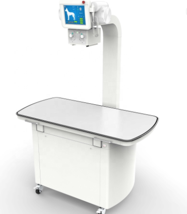
Veterinary Digital Radiography System
Product model:
DR-200Detailed introduction:
Veterinary Digital Radiography System (DR)
Model: DR-200 (1 6KW / 200 m A )
Features
★ Color LCD touch screen console, easy to operate.
★ Use 220V ordinary mains power supply, plug and play.
The high-frequency and high-voltage generator has the function of automatic calibration of the initial value of the tube current, which effectively prolongs the service life of the tube.
It has three modes of hand brake, foot brake and remote control exposure. The operator can perform exposure at a distance of ten meters, which greatly reduces ray radiation and effectively protects the health of veterinary personnel and animals.
The animal-specific photography bed is made of high-strength materials, which is waterproof, moisture-proof and corrosion-proof, with high X-ray transmission rate and clearer imaging.
★ The camera bed can be levitated and moved in four directions in front, back, left and right, which can better meet the needs of different animals for filming.Internationally renowned brand amorphous silicon flat panel detector, direct digital photography function allows excellent imagequality to be presented .
The side screen monitor is not only convenient for veterinarians to read pictures on the spot, but also greatly improves the grade of animal hospitals (optional).
The professional veterinary image workstation based on Windows operating system can directly print images without film, and can also realize remote consultation and case file storage.
Parameters
1. X-ray high frequency high voltage generator
1.1 Power supply voltage: single-phase three-wire system 220V±22V
1.2 Power frequency: 50Hz±1Hz
1.3 Nominal electrical power: 16 K W
★ 1.4 Maximum output power: 16 K W
1.5 Working frequency: ≥30kHz
★ 1.6 Tube current adjustment range: in the range of 16mA to 200mA, it can be adjusted in 12 grades
1.7 Adjustment range of tube voltage: continuous adjustment in the range of 40kV ~ 125kV, the adjustment step is 1kV
1.8 Exposure time range: 0.0025s~6.4s (2.5ms~6400ms)
1.9 Current time product range: 0.5mAs~320mAs
2. Microcomputer console
★2.1 Microcomputer console:color LCD touch screen
3. X-ray tube (Toshiba, Japan)
3.1 The highest peak tube voltage: 125kV
★ 3.2 Focus: Small focus: 1.0mm; Large focus: 2.0mm
3.3 Anode input power: small focus 21kW; large focus 42kW
3.4 Anode speed: 2700rpm
3.5 Target angle: 14°
3.6 Anode heat capacity: 140kHU
4. Beam limiter
4.1 Lead door control mode: manual
5. Photography bed
★ 5.1 Bed size: 1220x700x8 30 mm
5. 2 Maximum weight: 150 kg
★ 5. 3 bed surface movement: four-way floating
6. Flat panel detector (Korea cesium plate)
6.1 Material: amorphous silicon cesium iodide
6.2 Captured image size: 43×43cm (17×17 inches)
6.3 Pixel size: ≤127μm
6.4 A/D conversion: 16Bit
★ 6.5 pixel matrix: 3328×3328 (11 million pixels)
6.6 Preview time: ≤2 seconds
7. Image acquisition workstation
7.1 CPU: ≥2.8GHz
7.2 Memory: 4GB
7.3 Disk capacity: 1TB
7.4 Display: 23 inch LCD display
7.5 It can perform image acquisition, image zoom, image brightness adjustment, image contrast adjustment, image rotation, and image flip.
7.6 100M network interface; can carry out D ICOM 3.0 image transmission, and can query, edit, print, etc.
7.7 The DR system can manage patient animals and image information.

 2022-06-15
 hits
hits


